Showing posts with label DNA. Show all posts

Raymond Gosling: Flickr set dedicated to PhD thesis, 'X-ray diffraction studies of Deoxyribose Nucleic Acid '


Following the successful digitisation of Raymond Gosling's PhD thesis, 'X-ray diffraction studies of Deoxyribose Nucleic Acid' I have added a few of the images onto the project’s Flickr site: http://www.flickr.com/photos/51665752@N04/sets/72157631533614623/ .


(KDBP/5/1)


As I have already stated in the previous posts, Gosling was a vital worker in the King's effort to solve the structure of DNA. His PhD provides an excellent record of the experimental work going on at King’s - not only in terms of x-ray diffraction crystallography on the Signer DNA but the initial diffraction studies on sperm heads; the microscope-based work occurring simultaneously on nucleic acids and descriptions of model building, theoretic analysis of data and apparatus design are also described.


(KDBP/5/1)


The source, from an archival perspective, is ‘visually exciting’. Scientific records tend to be fairly cryptic with significance often hidden within the text or captured in a graph or table. The photographic prints within the thesis give a clear linear progression of the diffraction pattern that were being obtained at King’s. Broadly speaking, it is possible to see the improvement of images as the old x-ray equipment was discarded for the finer focus camera and x-ray tube and the presence of an expert crystallographer, in the form of Rosalind Franklin at the helm. 


Front cover of Raymond Gosling's PhD thesis (KDBP/5/1)


You can now access for free Raymond Gosling's thesis, via the Wellcome Digital Library- here. You can also search for other digital material relating to Raymond Gosling and other King's DNA workers via the Wellcome Library catalogue, as well as access collections from other archives relating to the history of modern genetics. 


Raymond Gosling: Visit to King's Archives


A few months ago, I had the pleasure of meeting Professor Raymond Gosling as he came into the Archives to be interviewed for a Swedish documentary on genetics. He was very talkative and full of insights into that crucial 1950-1953 period when the King’s Biophysics Unit were working on the structure of DNA. What was so refreshing about talking to him was not only his enthusiasm for the subject but his even-handed approach. Having worked with both Franklin and Wilkins at different stages of the project he gave a fair and independent analysis of their fractured relationship.

In the discussion with Gosling he emphasised the significance of the early x-ray photographs in kick-starting the hunt for the structure, and unintentionally spurring on the ‘race’ with its public unveiling at the Naples conference (1951). As well as describing the impressive character of the Biophysics Unit pioneered by Sir John Randall, he was aware that the time was ripe for the discovery of the structure of DNA and that several institutions around the world could have claimed it.


Below, is a video clip of Professor Gosling taking about the discovery of DNA at King's in the 1950s, as part of the London Science Festival, on the 21st October 2011. 


The diffraction pattern that Gosling refers to in the video is not the one shown in the video, which for those eagle-eyed readers out there is 'Photo 51'. Gosling was discussing the 1950 multi-fibre crystalline DNA pattern he produced with Wilkins and was the image shown at the Naples Conference in May 1951. Here is the correct image: 


X-ray diffraction image of DNA using the Raymax tube and Unicam camera, 1950 (ref: K/PP178/6/5/1)

The research papers relating to Raymond Gosling's work on DNA can now be accessed online via the Wellcome Digital Library. You can find out what other material available online via the Wellcome Library catalogue

Raymond Gosling: Not just an extraordinary envoy


Raymond Gosling was one of the key workers on DNA at King's during the period that became immortalised as the 'Race for the Double Helix'. His achievements have been eclipsed by the personality clash between Rosalind Franklin and Maurice Wilkins.  Gosling’s own role as the rift developed became that of "an Envoy Extraordinary and diplomatically carrying the "sense" of messages between them".  However, there was much more to Gosling's role than acting as a courier between the two parties and the next three posts will be dedicated to his role at King's. 

In the papers of Maurice Wilkins and those of the department of Biophysics Department are correspondence, articles, a PhD thesis, photographs and the diffraction patterns that Gosling produced with both Wilkins and Franklin. All these images have been selected for digitisation and this post contains a few samples of the type of material the collection holds. 

 One of our oldest pieces is a postcard sent to Wilkins from Gosling while on his honeymoon in 1950. His honeymoon consisted of touring Italy on his motorbike. Recently, I discovered that Wilkins, too, used to get lifts on the back of his motorbike after late nights working on x-ray diffraction experiments in the summer of 1950. Wilkins described this as “rather scary” but “enjoyed his hilarious time together” with Gosling. 


(ref: K/PP178/3/12)

(K/PP178/3/12)


The experimental biophysics work that King's has become known for in the solving of the structure of DNA was X-ray diffraction and Gosling's importance is undisputed. Gosling was the first to apply x-ray diffraction techniques on nucleic material at King's with the study of ram sperm heads in 1950. Later that year Gosling moved on to looking at DNA specimens and Wilkins brought him the calf thymus DNA donated by Rudolf Signer. The two began to collaborate and managed through innovation and some luck to produce a crystalline multi-fibre diffraction pattern of DNA that proved to be the breakthrough required to show that the structure of DNA was a feasible proposition. Soon afterwards new equipment and an expert crystallographer, in the shape of Rosalind Franklin, were brought in and Gosling was handed over to be Franklin's assistant. Gosling and Franklin made great strides and soon discovered through Franklin's control of the humidity levels that DNA occurred in two phases, called Structure A and B, and that this change could be reversible. This observation was followed up with further deductions based on the effect of water content on both structures. Whilst the rift within the lab affected joint efforts, Gosling and Franklin did go on to produce a three dimensional Patterson function of Structure A DNA, the data from which helped to prove the correctness of the Watson-Crick model.


Early calf thymus x-ray diffraction pattern produced by Raymond Gosling, 1950 (ref: K/PP178/5/1/1 Copyright: King's College London)


X-ray diffraction patterns of Structure A and B from Franklin & Gosling's article in Acta  Crystallographica,The  Structure  of  Sodium   Thymonucleate  Fibres. I.
The  Influence  of Water  Content', 1953 (ref: K/PP178/2/28 copyright: Please contact King's College London Archive Service)


  In the late 1970s, at the request of Wilkins, Gosling produced a recollection of his time at King's to allow Wilkins to reconstruct a popular account of DNA work at King's to counteract claims made by the recently published biography of Rosalind Franklin, by Anne Sayre (1975).

Some Recollections of DNA Studies in the Biophysics Laboratories at King's College. (ref: KPP178/5/6)

He described the atmosphere at King's as "friendly and positive". The interdisciplinary subject of biophysics was excitingly innovative and Gosling describes being 'exhilarated' by the prospect of investigating the 'dynamics of the single cell'


Some Recollections of DNA Studies in the Biophysics Laboratories at King's College.(ref: KPP178/5/6)


For Gosling, this period at King's could be broken down into four stages: firstly, the preliminary work around DNA before x-ray diffraction studies began and culminated in the first good crystalline pattern; secondly, the arrival of Franklin and the observation of two phases , Structure A and B, in DNA; thirdly, the interpretation of water content within the specimen and finally the ‘schism’ between Franklin and Wilkins.  


 In his concluding remarks, Gosling notes that it was his belief that Franklin never wholeheartedly believed that the structure was other than helical, despite giving that impression to Wilkins and Stokes. Perhaps the most telling observation is the following:

"If only Rosalind and Maurice had not been such shy people they might have been able to cooperate well in spite of their different views on how to set about solving the structure of DNA". 


You can read the full account of Raymond Gosling's reflections on his DNA work at King's via the Wellcome Digital Library- here


Digitising X-rays: the digitisation of the Biophysics collection


In this week’s post, I discuss some of the digitisation aspects of the project with special reference to the work of one of our digitisation contractors, MAX (previously MAX COMMUNCATIONS,) who digitised 4,000 of the glass and acetate material.

In October 2011, two of the digitisers from Max Ltd visited the archives in order to familiarise themselves with the collection and carry out some test scanning. The material was quite diverse: photographic prints, x-ray acetates, various sizes of glass plate negatives, folded negative rolls and 35mm mounted slides. The majority of this material required external specialists with the required expertise and equipment to undertake the scanning. A small test file was created composed of quarter plate glass negatives and x-ray acetates and sent to the Wellcome for approval. Both Iain Stringer, who would go on to digitise the collection, and David Cordery, head of Max Ltd, have prior professional experience of working with glass plate and acetate x-ray collections at various institutions  around the UK and we were fully confident of their ability to handle the fragile items and successfully scan them. The test images that were sent to the Wellcome were approved and scanning commenced in December 2011.

 The images were scanned at 300 dpi (dots per inch) at 8-bit (bit rate) RGB (Red Green Blue) using an Epsom V750 Pro scanner. This type of flatbed scanner is reliable and fast and from a preservation perspective, the scanner was suitable for digitising the x-ray acetates as the two- inch gap between the bed of the scanner and the top scanner head meant that it did not press onto the x-rays and so would not cause any further damage to the x-rays afflicted with vinegar syndrome (vinegar syndrome occurs when an acetate degrades and begins to oxidise creating a vinegar smell. The surface often begins to warp and crack and this eventually affects the emulsion. Unfortunately the process is irreversible and it is why digitisation is one of the most effective ways of preserving an accessible copy of the item.)

An example of an X-ray acetate diffraction image from the collection.  The  sleeve caption information has been added to the image. This diffraction image was taken by Wilkins around 1953-1955 and the likely source of the DNA originated from human subjects supplied by Leonard Hamilton and Ralph Barclay of the Sloan Kettering Cancer Center in New York.


I asked Iain to tell me about his experience scanning our material compared to his previous experience with similar collections. He said that the plates, in terms of general condition, were some of the best that he has worked with as hardly any were chipped or broken. The only slight issue that he encountered was that some of the slides were mounted with red strips, the adhesive of which had begun to seep and caused them to attach themselves to their transparent sleeves. In such cases, he therefore had to carefully remove the slide from its sleeve. This required a degree of perseverance, depending on the age and location of the adhesive strips on the slide.  

Regarding the x-ray acetates, I had assumed that this material would be trickier to scan considering the conditions that some of them were in. Iain surprised me by saying that for the purposes of scanning they were quicker to scan than the glass plates. Whilst care had to be taken in handling small, fragile and brittle objects like deteriorating x-ray acetates, the most time consuming element of scanning an x-ray was the post-production.

MAX Ltd provided us with images in three formats:  the raw TIFF original file, the enhanced TIFF amended file and a JPEG file. While enhancing an image can be difficult with regard to obtaining an authentic copy of the original, in a situation where the original is difficult to discern, post-production ‘clean up’ is necessary. The majority of the x-ray acetates retained a degree of visible content and by using Adobe Photoshop post production, it made it easier to enhance the original pattern of the x-ray and compensate for some of the surface damage caused by any deterioration.

Finally, I asked Iain what he thought of the acetate and glass material as a whole. He said:

“ I found the material quite interesting…I’ve learnt more about DNA than I have since school,, good thing about my job that I don’t have to concentrate on one specific thing. X-rays of DNA, diffraction, very interesting. They would definitely make a good print, stretched over a canvas, especially one of the really clear ones like ‘Photo 51’”

I agreed, diffraction patterns such as ‘Photo 51’ are visually striking though I personally am more in awe about the crystalline A-form DNA pictures as there is something rather mesmerising about the symmetry of these patterns. You can judge for yourself however, as these two x-ray patterns are shown below.


A-form DNA

B-form DNA

Will the archives of the future be made of the strand of DNA?

The prospect of combining archives and DNA feels like a plotline of a Twilight Zone episode. What's exciting however is that it is a distinct possibility. The Guardian have just released a story about the DNA inscription of a book (  http://www.guardian.co.uk/science/2012/aug/16/book-written-dna-code) initially reported in the US journal, Science. The book composed of 53,000 words includes eleven images and a computer program. The 5.27 megabit collection of data created over several days was produced by Professor George Church of Harvard Medical School.

The method they used, in principle, was the same as digital inscription: encoding all the book information into a binary sequence. The DNA base pairs in this case representing 1's and 0's with Arginine (‘A’) and Cytosine (‘C’) representing zero, and Guanine (‘G’) and Tyrosine (‘T’) representing one. The team developed a system in which an inkjet printer embeds short fragments of artificially synthesized DNA onto a glass chip. Each DNA fragment contains a digital address code that denotes location within the original fiDNAle.

What makes DNA such a brilliant medium for storage is its data storage with estimates suggesting a gram of DNA can store 455 billion gigabytes. The data is easily readable and copied and maintains its stability for several thousand years.  

The possibilities are fantastic. To put this in perspective, most digital formats require an upgrade after five years with physical data storage such as DVDs having, at most, a twenty year life span. This is because of the constant change in informational software packages within sturdy digital formats, such as TIFFs and PDFs having 10-15 year maximum life span. A DNA code sequence is therefore more desirable than a digital approximate, but neverless it is an exciting development and will potentially rival the paper record revolution in record keeping. This is an exciting archival perspective.

As a cataloguer and digitiser of the DNA related material of the Kings college London archive such a development is one of a personal joy. It would feel wonderfully apt to have the papers charting the discovery of the structure of DNA are encoded into DNA for future generations.

So long and thanks for all the hits!

  Sadly, this will be the last post on the DNA and Social Responsibility blog as the project is nearing an end. I hope those who have read the blog have taken away an interest in the life and career of Maurice Wilkins and how the papers that are held in the King's College Archive are a fantastic resource for future research in not only the history of genetics but also the wider role played by science in society in the twentieth century. Helping to catalogue this collection has been an enjoyable experience as it has introduced me to the delights of x-ray diffraction photographs; Fourier Transforms and Electron Density Maps not to mention the myriad political scientific groups most notably, BSSRS.

As a finale,I would like to sign off by sharing my favourite Wilkins' laboratory doodle. Cartoon is somewhat anarchic but does convey how science for Maurice Wilkins is a creative enterprise that still retained a slight hint of alchemy in the proceedings. 

Objectifying DNA

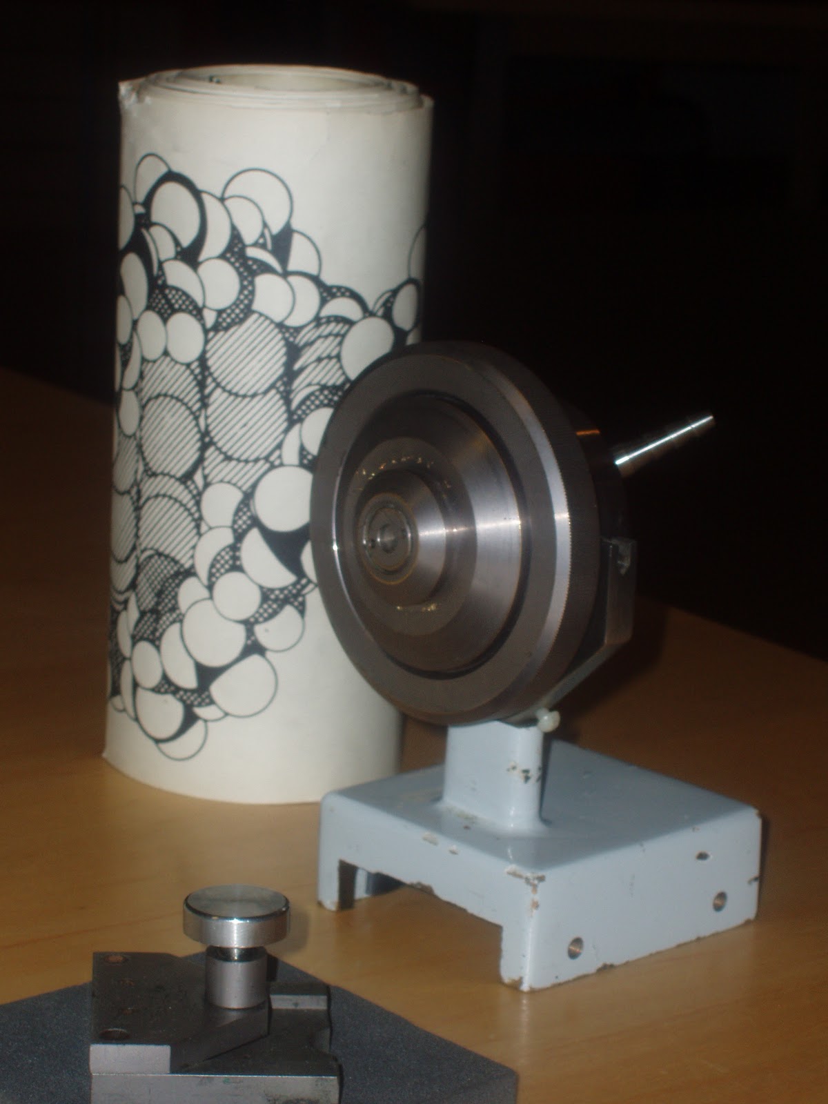
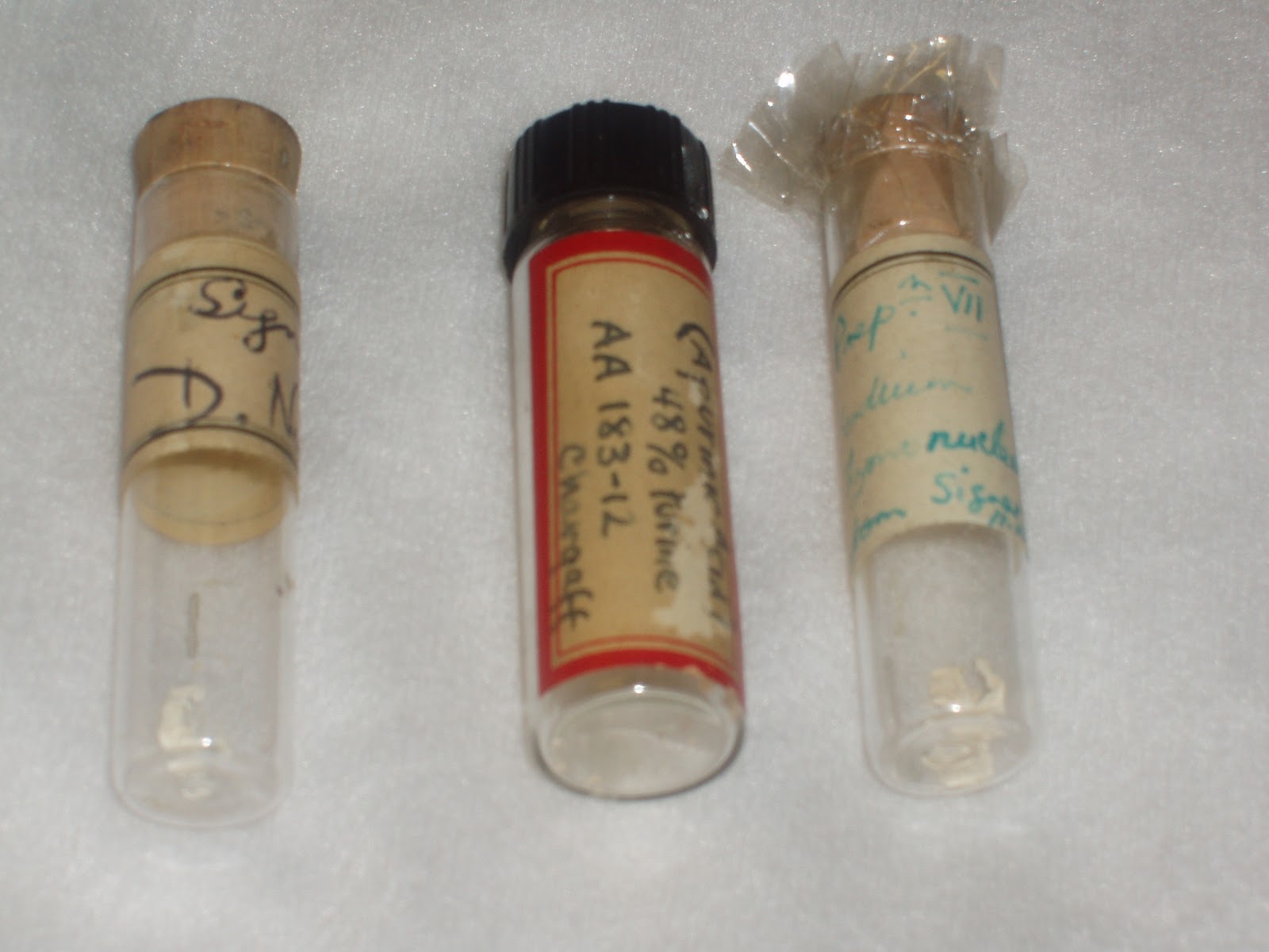
  The artefacts within the combined papers of the Maurice Wilkins and Biophysics collection include some of the most striking items in our holdings. Correspondence may have warmth and wit and the experimental notebooks may actually give the how and why but nothing beats the sheer visual punch of the DNA wire model or a X-ray diffraction camera. The artefacts are all the more impressive due to the diversity that the collection holds. Highlights include: original DNA fibres supplied by the Swiss biochemist, Rudolf Signer, which were the main source of DNA used in the X-ray diffraction experiments by Rosalind Franklin and Raymond Gosling; several X-ray diffraction cameras including the micro camera used to obtain "Photo 51", the picture of B structure DNA that so sharply showed a helical structure of DNA; DNA models and diagrams that the Biophysics Unit used to construct and refine more detailed models of DNA, with the pride and joy being the several metre long roll of the DNA molecular model ceremoniously nicknamed the "DNA toilet paper". These are a few of our favourite things and they are joined by many other items that directly relate to DNA and previous microscopic research carried out on the subject.


A few of the DNA related artefacts at King's Archives such as the single fibre X-ray camera used by Rosalind Franklin, glass vials containing original DNA samples used in the X-ray diffraction experiments, DNA diagram and Biophysics photo index


The collection has a remarkable degree of preserved DNA samples from the 1950s that not only include the Rudolf Signer Calf Thymus DNA that produced excellent B configuration X ray patterns sample but also DNA samples prepared from other scientists such as Erwin Chargaff and Leonard Hamilton which used a variety of other DNA sources like bacterial cultures and human DNA.

Image of a molecular model of DNA reproduced for the 40th anniversaryof the discovery of the double helix at King's College London

Two new Flickr sets taken from 35mm mounted slide series. Only a small sample of over thousand images that were digitally captured over the last two months. Highlights include high quality images of Rosalind Franklin, x-ray diffraction images and models of DNA and images relating to Maurice Wilkins' involvement in the Campaign for Nuclear Disarmament.

"Dear M": the transatlantic DNA collaboration of Maurice Wilkins and Leonard Hamilton

This week's post calls attention to a series of correspondence written in the 1950s that documents the DNA research carried by Wilkins and his group at King's College London from 1953 to1960. The correspondence is with Dr Leonard D Hamilton, a British medical researcher and pathologist based in the United States who worked for the Sloan Kettering Institute for Cancer Research and later the Brookhaven National Laboratory. He provided DNA samples to Wilkins at King's from 1952 into the 1960s. As my title suggests the two were firm friends and their letters are refreshingly candid. Although these letters do not document the breakthrough research on the structure of the double helix like the similar correspondence between Wilkins and Crick they do give us a good picture of how Wilkins coped with the news and life thereafter.


DNA Samples: Supply and Demand

In 1953, Leonard Hamilton became the principal provider of DNA for the King's team. Samples had previously been provided by a host of other international scientists such as Rudolf Signer, Erwin Chargaff and Harriet Ephrussi and there was a great pressure to produce better DNA samples now it had been established that DNA was the basis of genes.Wilkins and his team were also under pressure to provide a more detailed double helical model to confirm that the structure was correct and not an equally valid alternative structure.

Figure 1: Photocopy of a letter from Wilkins to Hamilton, dated 28 May 1953: Wilkins thanks Hamilton for the DNA sample taken from a mouse sarcoma and generally expresses an eagerness to press on with further DNA research.

 In the above letter, Wilkins shows his excitement over the diffraction image obtained using the S-180 mouse sarcoma sample. He also expresses the need for greater quantities and variant salts and solutions that becomes a constant theme of this correspondence, as this quotation from a letter from Wilkins on the 9 June 1954 reveals:

"As I thought I made clear, what we need is better DNAs and hence I was very disappointed to find all the samples you sent recently were no better than the usual quality. We have not the time to test laboriously numerous samples in the hope one might be good. The three dry samples were have had already and those in solution, owning to a lack of warning, arrived at this end and were held at the airport for several days and appear to have deteriorated". 

The problem of the supply of demand of DNA were exacerbated by the trans-Atlantic nature of the partnership. Communication was the biggest issue. Before the days of global mass-communication, the main medium for communication was by letter or telegram (international telephone calls had only recently been introduced and were very expensive). The frustration was felt on both sides of the Atlantic especially with the additional pressures of journal and conference deadlines. Despite these stresses, they made progress as this quote from Wilkins on 5 November 1954 states:

"The 10 week exposure came off today and is a great success. it is one of the biggest improvements we have ever had in A pictures and is very much better than the best before. Will send a copy soon and I hope I will spur you on to even better things...Good old Leonard!"


Living in the Shadow of the Watson-Crick Model

One of the reasons why these letters make compelling reading is the scattered references to the discovery of the Double Helix. In the letters we get a contemporary commentary on the initial impact of the discovery and personal opinions on some of the key characters such as Francis Crick, James Watson and Rosalind Franklin. Of particular interest is the response of Hamilton who considered that Wilkins and King's College London Biophysics Unit had been hard done by Watson and Crick's model.


Figure 2: Letter from Leonard Hamilton to Wilkins dated October 12 1954: In this letter, Hamilton describes a visit from Rosalind Franklin and also his plan to air off a little "pro-Wilkins propaganda!" at a nucleic acids conference. 
 From the correspondence, it is clear that the battles and wounds caused by the discovery are still fresh but the scientific interest in the findings ( such as the journal Scientific American in Figure 2) are already calling for a narrative of the discovery. Hamilton's repeated opposition to calling the double helix the Watson-Crick model is an example of this increasing scientific interest and his own attempts to inform the American scientific community of the role that King's Biophysics Unit played in its creation. 

Wilkins' own initial thoughts and feelings were somewhat more ambigious. For example, his assessment of Francis Crick in the letter shown in Figure 1 states:

"Taken the Watson Crick model with a grain of salt. Francis is quite certain he can solve all the problems of the universe by pure thought and his lack of facts had lost him many friends temporarily until he recovers from DNA hysteria. I am very fond of Francis but he can be a bit much at times and he can be a ruthless careerist when he thinks it suits him. but keep that quiet to ourselves. We both like him a lot so there is no harm saying this to you".

His reaction is in line with his famous letter to Crick on hearing the discovery where he called Crick and Watson a couple of "old rogues" but accepted the model gracefully. Yet in this letter he does admit a degree of resentment over the Crick's "careerist" tendencies but not enough to irreprecibly damage their friendship. 


Finals thoughts

The correspondence between these two friends is of great reading for anyone interested in the story of the double helix and the DNA research carried out at King's College London. By being both a commentary and account of the DNA research undertaken at King's in the fifties the letters acts as an informal guide to the work being carried out and excellent source of information of the different DNA salts and samples. For me, however, the best aspect of the letters is the openess and informality that Wilkins shares with his friend about his life and research.  

Figure 3: Leonard Hamilton and Maurice Wilkins together in the 1960s.


Final words

"Do please reply soon. The nature of the gene depends on it. What piffle. M"

DNA on Display: Strandlines community visits King's Archive

 On 23 February 2011, King's College London Archives hosted a local event organised by the Strandlines Digital Community. A diverse group spent the afternoon exploring many of the treasures of our archives including artifacts and documents from our DNA collection. Information and anecdotes about the collection were provided by Patricia Methven, Head of King's College Archives and senior archivist Geoff Browell. A project blog on the Strandlines website provides a more comprehensive coverage of the day plus feedback from a number of visitors (http://www.strandlines.net/blog/archives-afternoon-23rd-february-2011).

Strandlines event at King's College London archives on the 23 February 2011


As the DNA & Social Responsibility project assistant, I was delighted to see the level of interest and enthusiasm for the DNA collection, whether it was copies of 'Photo 51' or the 'wire model of DNA'. Not only were people awe-struck by the beauty and importance of DNA artefacts and photographs but they were also charmed by the more personal items of our collection such as 'Radium Island', the boyhood adventure story by Maurice Wilkins. Overall, it was good to see that visitors come away appreciating the long tradition of scientific innovation at King's and the beneficial role that the archives can play in the local community.

Short and Simple (ish) Guide to X-ray Diffraction


  X-ray diffraction (or X-ray crystallography) was the chief physical method used to determine the structure of DNA. In this post, I will briefly and as simply as I can (which with my non-scientific background should not be a problem!) explain what x-ray diffraction technique is and its relative importance to the overall discovery.


What does X-ray diffraction actually mean?
 X-ray diffraction is the method of projecting a beam of X-ray radiation at a target object and through to a photographic film on the far side. A series of spots appear on the photographic film following this exposure, which is formed by the x-ray radiation diffracting off the structure that they passed through. These diffraction patterns give an indication of the general structure of the object (such as an inorganic crystal or macro- molecule such as DNA) which can then be delineated using complex mathematical formulas.

Why use X-rays in the first place?
The reason why X-ray beam is required in the first place is that atoms are too small (0.1nm between them, bearing in mind that 1 millimetre = 1000000 nanometres) to be revealed using visible light and therefore could not be viewed by a light microscope (even an electron microscope does not possess the required magnification). X-ray radiation fits the appropriate wavelength to be diffracted by the object and produce visible results.   

What causes the diffraction of the X-ray beams?
What the X-ray beam are diffracting is not the entire atom but the orbiting electrons (one of the component parts of an atom) that are close enough to the core (nucleus) of the atom to give a good indication of the structure of the unit cell (the term used for the repeating unit found in crystals and macromolecules). The end image is known as an electron density map of that unit cell. However due to the incredibly weak image a single molecule would produce, a crystalline structure is used instead, for example common salt (NaCl), since a crystalline structure provides a huge number of molecules arranged in the same orientation and therefore produces the same scattering effect on the X-ray beams. 

In this diagram, the diffraction of the X-ray beam causes an image with a helical arrangement to form as all the DNA molecules in a fibre are aligned in the same direction.


X-ray diffraction of nucleic acids at King’s College London from 1950 to 1953 
 X-ray diffraction studies on DNA began in June 1950 when Maurice Wilkins asked PhD student Raymond Gosling to assist him in diffracting the DNA fibre samples prepared by the Swiss biochemist, Rudolf Signer. Fibre diffraction did not usually provide good quality images because of the thinness of the fibres and therefore a very small mass to scatter the radiation. Nevertheless, the fibres’ remarkable uniformity when wetted allowed Wilkins to manipulate them into a bundle and mount them on a wire frame to obtain x-ray diffraction images. The initial images showed promise but Wilkins and Gosling were greatly assisted by J T Randall’s own experience with X-ray diffraction.  He advised how the surrounding air could affect the x-ray scattering. The solution was to pass hydrogen through the camera and control the relative humidity of the sample.  With this in place, the resulting images were much sharper and showed a clear crystalline diffraction pattern.
X-ray diffraction pattern obtained by M H F Wilkins and R Gosling in late 1950 showing a clear crystalline arrangement.


It was in late 1950 that the theoretical physicist Alec Stokes first noticed an interesting observation from the images. He realised that there was no diffraction at all along the length of the molecules: a sign that DNA might be helical.  However, the King’s College team needed far sharper images to confirm this hypothesis. This required a new X-ray camera that could work on single fibres.  Through a fortunate coincidence, Werner Ehrenberg and W E Spears had just developed one at Birkbeck: this was generously loaned to the King’s College team.

Before the new camera was set up, it was decided that Rosalind Franklin, who was joining the laboratory from Paris, would replace Wilkins in producing the x-ray diffraction images with the continued assistance of Raymond Gosling. Both Stokes and Wilkins continued working on the problem with the latter embarking on some rough tests with the old X-ray diffraction camera on various DNA specimens that produced an observed “X” crossed pattern. The X pattern of diffraction was created by the x-ray radiation scattering at right angles off the "zigzag" structure of the DNA chain.  This interpretation was further supported when Franklin and Gosling produced the first “B” structure X-ray patterns in the late summer of 1951. This was a crucial development as it showed two observed states of DNA: crystalline “A” and semi-crystalline “B” (the best B structure diffraction photograph became known as “Photo 51”). The photos also supported the predicted observed readings of a helix that Alec Stokes had developed using the mathematical technique known as Bessel functions.
Plot of Bessel Functions for a smooth helix, named "Waves at Bessel-on-sea" by Alec Stokes who completed the calculations for the diagram over a single train journey.


It was now Maurice Wilkins and Rosalind Franklin disagreed over the direction of the research in finding the overall structure. Wilkins was keen on hypothetical model building while Franklin favoured a more systematic study of the structure. This parting of ways can be partially explained as stemming from the limitations of the x-ray diffraction process itself. For example, the evidence from the photos clearly pointed to a helical structure but this begged question: what type of helix? Helices in nature could occur in single, double and even triple strands and there was no clear indication, which was the right number. This is why the King’s College London attempt at model building proved to be a failure when the model made by Bruce Fraser showed a triple helix based on the chemical readings but was unable to fit with the rest of the x-ray data. A crucial piece of the puzzle was missing and related closely to DNA’s function of providing the genetic material for life: it was only when Jim Watson and Francis Crick came up with the base pair hypothesis that the double helix seemed the ideal form. 


In this diagram, we can see the general similarity between a single and a double helix.


X-ray diffraction studies undertaken at King's College London provided part of the experimental structural data needed to solve the general structure of the DNA double helix. Yet, as important as these observations were other methods and disciplines were of equal importance in unravelling the overall structure, in particular the biochemical work of Erwin Chargaff and the biological insight of Jim Watson. X-ray diffraction work on DNA at King’s did not finish with the unveiling of the structure in March 1953 but continued for another decade as Wilkins and his team worked to test to the correctness of the "Watson-Crick" model. 


Public unveiling of frieze celebrating Rosalind Franklin and Maurice Wilkins work at King's College London


On the 13th September 2010 Principal Professor Rick Trainor unveiled the newly designed DNA frieze outside the Franklin-Wilkins building on the Waterloo Campus. The new permanent window display was sponsored by Ecovert FM to mark the 10th anniversary of the Public Private Partnership (PPP) contract with King's.  The set of friezes depict the two scientists and the key developments associated with them that led to the solving of the structure of DNA. To celebrate the unveiling members of the Franklin and Wilkins family joined the Principal and the CEO of Ecovert Group Bruno Bodin for this special occasion.


(Left to right) Jenifer Glyn, her husband Ian, Sarah Wilkins, CEO of Ecovert Group Bruno Bodin, George Wilkins, King's Principal Professor Rick Trainor and at the front, George Wilkins' two sons.


The set of friezes on street level on Stamford Street give an excellent introduction to the DNA work undertaken at King's: for the representation of Rosalind Franklin it praises the X-ray diffraction studies that she and Ray Gosling took and couples her with her most famous creation, 'Photo 51'. It's visually arresting for the casual pedestrian or distracted student but the real treat is the detail on the friezes: the extracts and images of Franklin's notebook and a brief diagrammatic explanation of the significance of 'Photo 51'. 

Frieze panel of 'Photo 51' with explanation below. 


 Maurice Wilkins the frieze acknowledges both the work that he achieved in the early fifties and the later work verifying the Watson-Crick model. Again the frieze is a fine tribute pointing out the early X-ray diffraction work and early helical interpretations harbored by Maurice Wilkins and his collaborator Alec Stokes. 


 Alongside these panels on the story of DNA is the impressive representation of the scale and enormity of DNA through a linear outline of a section of a double helix coiled around the three revolving doors of the entrance of the Franklin-Wilkins building. The creator of the artwork, Ian Chilvers of Atelier Works, aided by Dr Roland Roberts of the Department of Medical & Molecular Genetics here at King's College, scaled up a section of DNA from the smallest human chromosome (21) by a factor of 1.1. billion and applied the design to the doors used a frosted linear vinyl. The artwork gives a visual demonstration of the complexity of our genetic make-up: In order to depict the total size of this chromosome at this scale it would involve having to stack 10 million of these doors on top of each other- making it a staggering 20,000 km high!

Model of the DNA design courtesy of Atelier Works
The project is part of a larger renovation scheme by KCL and its facilities management partner Ecovert FM to reduce the ecological impact of all campus buildings. The Franklin-Wilkins building can now harvest rainwater for cleaning and toilets, reduce energy waste through energy efficient lighting and heating systems and even convert waste into biomass. This larger project would have pleased Maurice Wilkins who was a keen advocate of alternative energy use and would appreciate that a building that shares his name was pursuing an innovative and environmentally responsible policy towards energy consumption.

Exterior of the Franklin-Wilkins building on Stamford Street